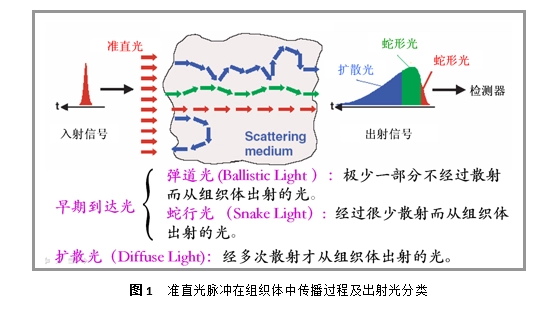
Biological tissues (e.g., skin, muscle, etc.) absorb and scatter both visible and near-infrared light, and are therefore a “turbid medium”. Light through the turbid medium, in the photon is absorbed by the medium or by the medium before the emission, its moving trajectory in the medium is random, some photons in the direction of its movement scattered less, so it will be close to the direction of its incidence trajectory from the medium, known as ballistic light (Ballistic Light). Some photons are scattered many times and their trajectory is very random and irregular, called Diffuse Light. Since biological tissues are high scatterers, diffuse light usually dominates the light emitted from biological tissues, and ballistic light only accounts for a small portion of the light, and as the sample becomes thicker, the proportion of ballistic light will be further reduced. Therefore, in recent years, light scattering with diffuse light as the research object has gradually become a research hotspot. Combining optical imaging means and existing medical imaging means such as CT, MRI, etc. can greatly improve the safety, nondestructiveness and accuracy of clinical biomedical imaging technology. Diffuse optical tomography (DOT) is a technique that uses diffuse light to obtain tissue information.
Translated with DeepL.com (free version)
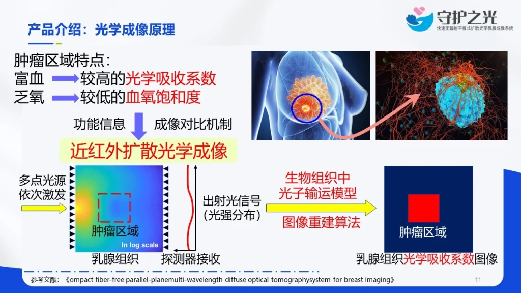
DOT provides a new technical means for non-destructive detection based on tissues, using the optical properties of the tissues it provides we can identify and diagnose the physiological state of the measured tissues, such as normal tissues, cancerous tissues, diseased tissues and other tissue states, so as to realize the early diagnosis of diseases, which is of great practical significance and practical value in the application of clinical medicine. Currently, DOT has been applied to the clinical diagnosis of many diseases, such as the measurement of blood oxygen saturation in the brain of newborns, the detection and imaging of breast cancer, the imaging of tumors, and the functional imaging of the cerebral cortex. It is believed that with the further development and maturity of this technology, the areas of its application will also increase.
Translated with DeepL.com (free version)