
Lab Introduction
Facing the “Healthy China 2030”, the localization of medical equipment and other major national strategic needs, clinical needs and biomedical frontier, combined with the strong electronic information background of Xi'an University of Electronic Science and Technology, the team is committed to the research and development of highly sensitive, high specificity, high spatial resolution multimodal imaging systems and imaging technology, for the early stage of disease, accurate diagnosis and efficacy assessment and basic research in life sciences to provide powerful imaging technology means! We are committed to developing highly sensitive, high spatial and temporal resolution multimodal imaging systems and imaging technologies to provide powerful imaging technology for early disease diagnosis, efficacy assessment and basic life science research!
News
Upcoming Lab Events
- There are no upcoming events.
research direction
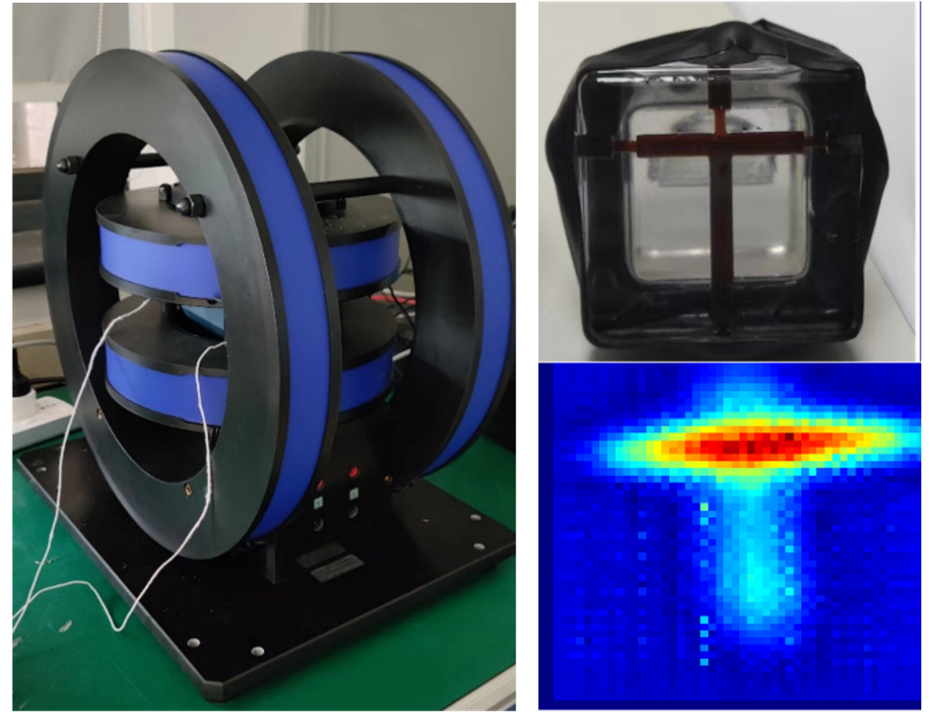
MPI
Magnetic Particle Imaging (MPI) is a tracer method for detecting the spatial distribution of magnetic nanoparticles based on functional and tomographic imaging techniques. As functional imaging, MPI improves imaging resolution without the use of radioactive material, making it the most clinically translatable technique in the last 20 years.
Micro-CT
Micro-CT (micro computed tomography), also known as micro-CT, micro-focused CT or micro-CT, is a technology that employs a micro-focused X-ray bulb, which is different from ordinary clinical CT, to scan and image live small animals or a variety of hard tissues and related soft tissues with a resolution of up to a few micrometers, second only to the level of simultaneous accelerated X-ray imaging equipment, with a good “micro” effect. It has a resolution of several micrometers, which is second only to the level of synchronized accelerated X-ray imaging equipment, and has a good “microscopic” effect.
Cherenkov radiation imaging
Cherenkov radiation is a continuous-spectrum visible radiation produced by high-energy charged particles moving at more than the speed of light in a dielectric medium. In addition to detecting charged particles in high-energy physics, Cherenkov radiation can also be used to image radionuclide-labeled drugs and radiation therapy rays for clinical diagnostic applications in biological research.
PET
PET uses annihilation radiation and positron collimation (or photon collimation) technology to non-destructively, quantitatively, and dynamically determine the spatial distribution, quantity, and dynamic changes of PET contrast agent or its metabolite molecules in vivo, and to obtain biochemical, physiological, and functional metabolic information on the interactions between PET contrast agent and its targets (e.g., receptors, enzymes, ion channels, antigenic determinants, and nucleic acids) in vivo at a molecular level, to provide important information for clinical research. It provides important information for clinical research.
Photoacoustic Imaging
Photoacoustic Imaging (PAI) is a new non-invasive and non-ionizing biomedical imaging method developed in recent years. The photoacoustic signals generated by biological tissues carry information about the light absorption characteristics of the tissues, and by detecting the photoacoustic signals, an image of the light absorption distribution in the tissues can be reconstructed. Photoacoustic imaging combines the advantages of high selectivity in purely optical tissue imaging and deep penetration in purely ultrasound tissue imaging to obtain high-resolution and high-contrast tissue images.
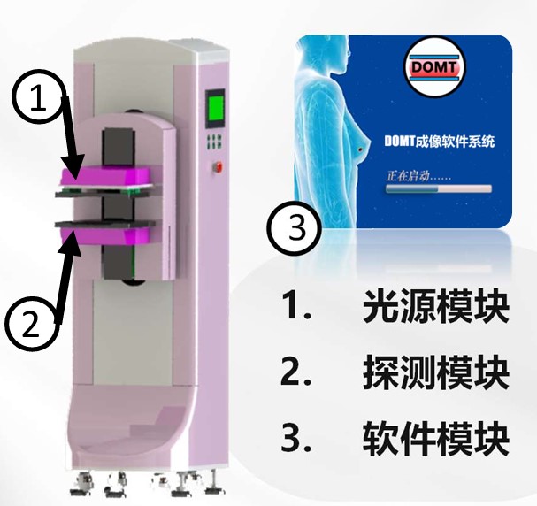
DOT
漫射光学层析成像(Diffuse Optical tomography,DOT)是一种面向厚组织体的利用近红外光(600-900nm)照射获得的三维功能成像方法,其目标是通过发展高灵敏的近红外光子检测仪器和基于生物组织光子输运模型的图像重建技术,从多点激励下表面扩散光的测量信息中反演组织体内部光学特性参数的三维分布,并使之与该组织的生理状态(血红蛋白浓度及氧饱和度等)相关联。
Achievement
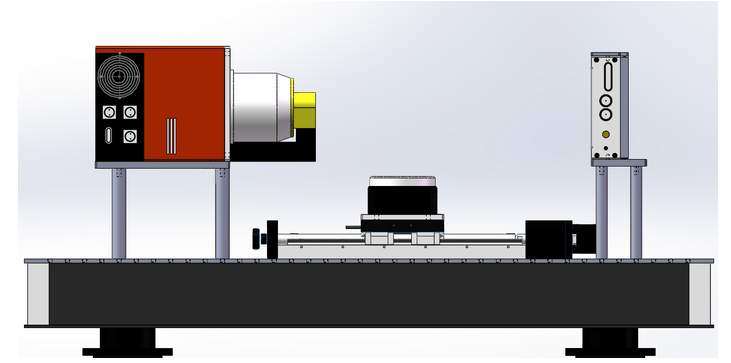
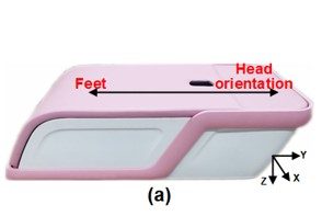
The team's research work involves micro-CT imaging, micro-PET imaging, photoacoustic imaging, diffuse optical imaging, Cherenkov imaging (CLI), optical projection tomography (OPT), X-ray luminescence tomography (XLT), and other imaging modalities, and the research content includes the development of imaging systems and imaging key technologies, imaging theory and reconstruction algorithms, GPU-based accelerated high-performance computing, medical image processing, multi-modality data fusion, medical software development, biological and clinical experimental validation, and other aspects. The research includes imaging system development and imaging key technology research, imaging theory and reconstruction algorithm research, GPU-accelerated high-performance computing, medical image processing, multimodal data fusion, medical software development, biological and clinical experiment validation, and other aspects. In addition, we have developed our own platforms and equipments in various directions.
导师团队

Prof. Shouping Zhu
Master's/Doctoral Supervisor, Xi'an University of Electronic Science and Technology
Prof. Xu Cao
Master's/Doctoral Supervisor, Xi'an University of Electronic Science and Technology
Associate Prof. Yihan Wang
Master's Degree Supervisor, Xi'an University of Electronic Science and Technology
任胜寒 讲师
Master's Degree Supervisor, Xi'an University of Electronic Science and Technology
刘相宇 讲师
西安电子科技大学博后